
Human eye diagram, Eye anatomy, Eye diagram
Labelling the eye Interactive Add to collection Use this interactive to label different parts of the human eye. Drag and drop the text labels onto the boxes next to the diagram. Selecting or hovering over a box will highlight each area in the diagram. Cornea Lens Retina Optic nerve Pupil Schlera Vitrous humour Iris Download Exercise Tweet
:max_bytes(150000):strip_icc()/eye-conjunctiva-871453538-5a26c6ad22fa3a0037d5edad.jpg)
How the Human Eye Works (Structure and Function)
Diagram of human eye anatomy with label illustration. Download a free preview or high-quality Adobe Illustrator (ai), EPS, PDF, SVG vectors and high-res JPEG and PNG images.

Brain Post How Big is Your Blind Spot? Human eye
Cornea of the eye Uvea of the eye Pupil: Aperture of the eye The retina: Where vision begins Macula lutea of the eye Choroid of the eye Lens of the eye Ciliary body Eye muscles Aqueous humor Optic nerve Fovea centralis Optic chiasm And for a description of common vision problems, see Refraction and refractive errors: how the eye sees.

Pin on Premed/med school stuff
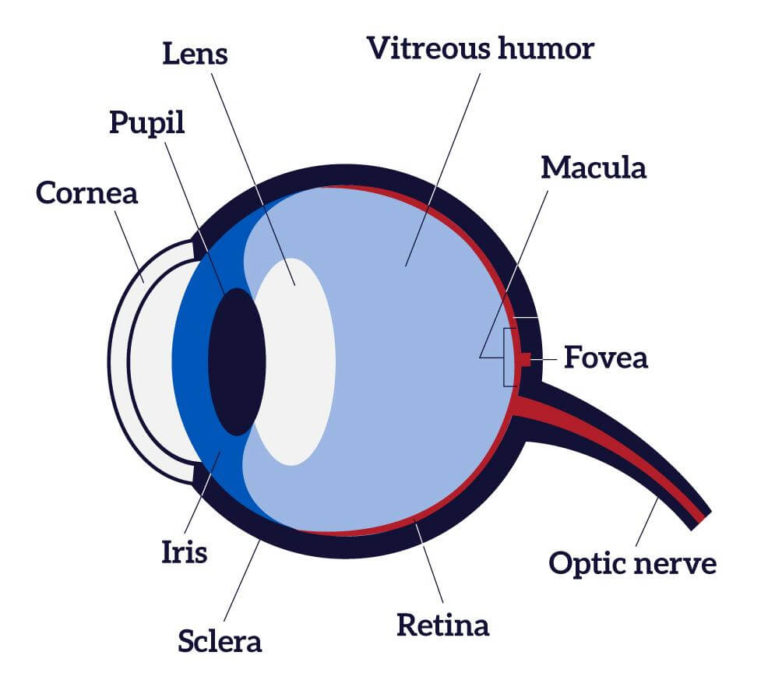
Take a look at the diagram of the eyeball above. Here you can see all of the main structures in this area. Spend some time reviewing the name and location of each one, then try to label the eye yourself - without peeking! - using the eye diagram (blank) below. Unlabeled diagram of the eye. Click below to download our free unlabeled diagram of.

OUR EYES WORK LIKE CAMERA’S! Discovery Eye Foundation
Human Eye Diagram: Contrary to popular belief, the eyes are not perfectly spherical; instead, it is made up of two separate segments fused together. Explore: Facts About The Eye To understand more in detail about our eye and how our eye functions, we need to look into the structure of the human eye. Recommended Video: 1,221

Human eye Extraocular Muscles Britannica
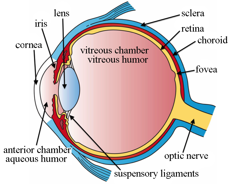
The diagram below points to different parts of the human eye. The human eye. Choose the correct labels for the parts shown. Choose all answers that apply: A is the crystalline lens. A A is the crystalline lens. B is the aqueous humour. B B is the aqueous humour. C is the iris. C C is the iris. D is the cornea. D D is the cornea.
Vision and Eye Diagram How We See
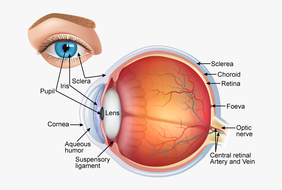
The light passing through cornea, pupil, and lens gets focused on the retinal membrane. In addition to tissue components, retina is made up of two types of cells: rod cells and cone cells. The.
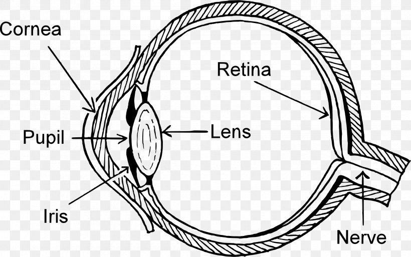
Diagram Human Eye Eye Pattern Clip Art, PNG, 2400x1503px, Watercolor
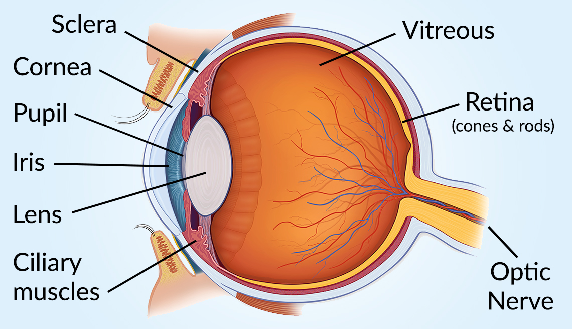
Eyes are approximately one inch in diameter. Pads of fat and the surrounding bones of the skull protect them. The eye has several major components: the cornea, pupil, lens, iris, retina, and sclera.

Eye Diagram Unlabelled Human Eye Diagram Unlabelled Human eye diagram
Cornea. The clear, dome-shaped surface that covers the front of the eye. Iris. The colored part of the eye. The iris is partly responsible for regulating the amount of light permitted to enter the eye. Lens (also called crystalline lens). The transparent structure inside the eye that focuses light rays onto the retina. Lower eyelid.

Labelled Diagram Of Human Eye , Png Download Label A Human Eye
1. Conjunctiva The conjunctiva is the membrane covering the sclera (white portion of your eye). The conjunctiva also covers the interior of your eyelids. Conjunctivitis, often known as pink eye, occurs when this thin membrane becomes inflamed or swollen. Other eye disorders that affect the conjunctiva include:
Human Eye Labelled Diagram , Free Transparent Clipart ClipartKey
Biology Article Diagram Of Eye Diagram Of Eye The human eye is responsible for the most important function of the human body, the sense of sight. It consists of several distinct parts that work in coordination with each other. The most common eye diseases include myopia, hypermetropia, glaucoma and cataract.
Draw a labeled diagram of the V.S. of the human eye and write one
Download. English: Parts of the Eye (PDF 603.5 KB) Spanish: Las partes del ojo (PDF 897.7 KB) Check out this fact sheet to see a labeled diagram of the eye and learn about the different parts of the eye.
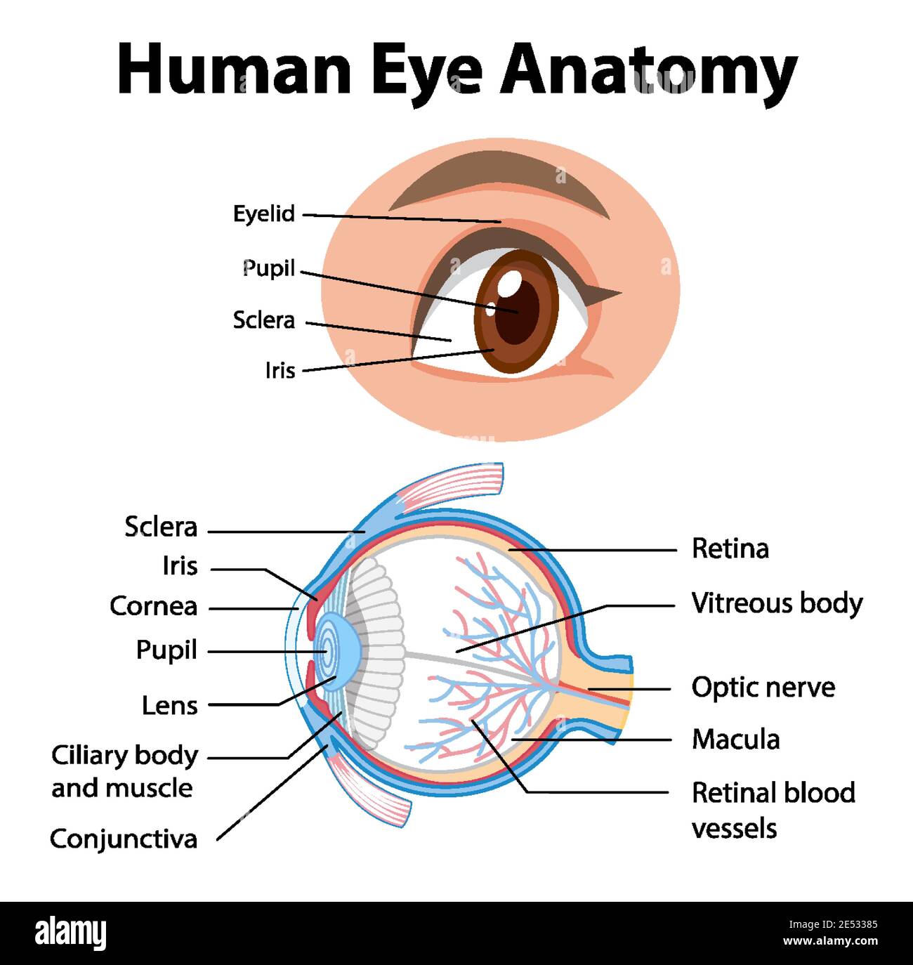
Diagram of human eye anatomy with label illustration Stock Vector Image
Anatomy of The Eye. The anatomy and physiology of the human eye is an important part of many courses e.g. in biology, human biology, physics, and practical courses in medicine, nursing, and therapies. This simple introduction the subjects of 'the eye' and 'visual optics' includes a simple diagram of the eye together with definitions of the.
Human Eye Diagram, How The Eye Work 15 Amazing Facts of Eye
human eye, in humans, specialized sense organ capable of receiving visual images, which are then carried to the brain.. Anatomy of the visual apparatus Structures auxiliary to the eye The orbit. The eye is protected from mechanical injury by being enclosed in a socket, or orbit, which is made up of portions of several of the bones of the skull to form a four-sided pyramid, the apex of which.
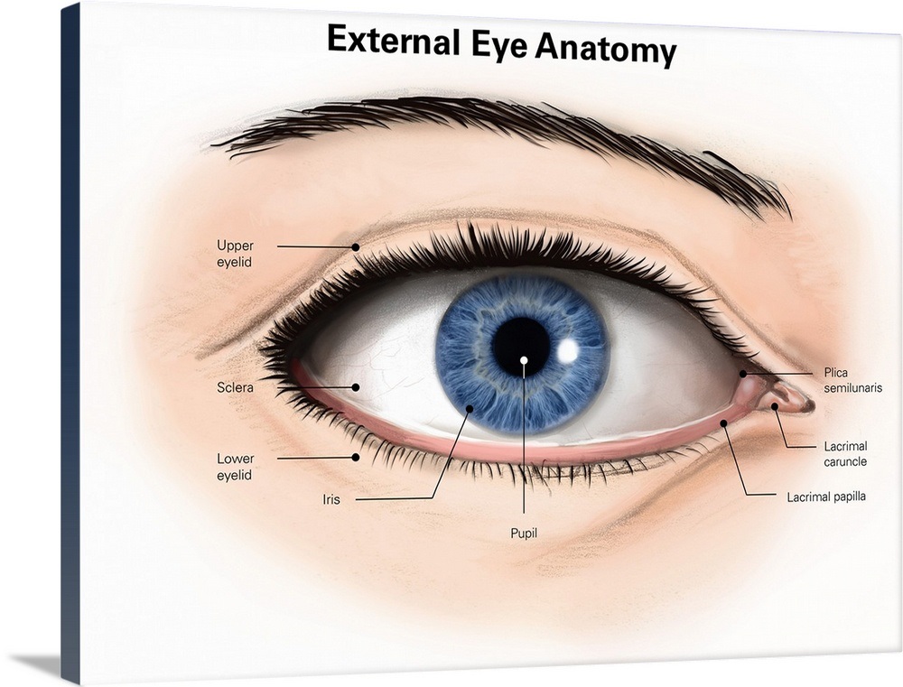
External anatomy of the human eye (with labels) Wall Art, Canvas Prints
Diagram of the Eye Posted in Eye Health, Uncategorized | August 5, 2018 Even though the eye is small, only about 1 inch in diameter, it serves a very important function - the sense of sight.
/GettyImages-695204442-b9320f82932c49bcac765167b95f4af6.jpg)
Structure and Function of the Human Eye
6 min read Your eye is a slightly asymmetrical globe, about an inch in diameter. The front part (what you see in the mirror) includes: Iris: the colored part Cornea: a clear dome over the iris.
